
Understanding Birdshot Chorioretinopathy: Causes, Treatments, and Real-Life Outcomes
Being told you have a rare eye disease like birdshot chorioretinopathy can feel terrifying—especially when you start to notice changes in your vision and realize there isn’t much awareness about it. If you or someone you love has recently received this diagnosis, you’re not alone in feeling overwhelmed, confused, and urgently in need of clear, trustworthy information.
In this guide, we’ll walk through what birdshot chorioretinopathy is, how it’s usually diagnosed, which treatments are commonly used as of 2026, and what the long‑term outlook can look like—with a focus on practical steps you can take to protect vision and quality of life.

What You’ll Learn in This Article
- What birdshot chorioretinopathy is and how it affects the eye
- Why it’s considered an autoimmune disease and its genetic link
- How doctors diagnose it and monitor progression
- Current treatment options and how they’re typically combined
- Realistic expectations for prognosis and day‑to‑day life
What Is Birdshot Chorioretinopathy?
Birdshot chorioretinopathy (often shortened to “birdshot” or “BCR”) is a rare, chronic autoimmune disease that affects two key structures at the back of the eye:
- Choroid: the layer of blood vessels that nourishes the retina.
- Retina: the light‑sensitive “film” lining the back of the eye, essential for vision.
Under the microscope and on specialized imaging, doctors see multiple pale, cream‑colored spots in the back of the eye that can resemble the pattern left by birdshot pellets—hence the name. Despite the dramatic appearance, the disease often starts subtly.
BCR is categorized as a type of posterior uveitis (inflammation at the back of the eye). It is:
- Bilateral: it almost always affects both eyes.
- Chronic: it tends to last for years and needs long‑term follow‑up.
- Progressive: without treatment, it can slowly damage vision.
An Autoimmune and Genetic Disease
Birdshot chorioretinopathy is considered an autoimmune disease. That means the immune system, which normally protects us from infections, mistakenly attacks the eye’s own tissues—particularly cells in the choroid and retina.
It is strongly associated with a genetic marker called HLA‑A29. Over 90% of people with birdshot carry this marker, compared with a much smaller percentage in the general population. However:
- Having HLA‑A29 does not mean you will definitely develop birdshot.
- Not everyone with birdshot will develop other autoimmune diseases.
“HLA‑A29 is one of the strongest genetic associations we see in uveitis, but it is only part of the story. Environmental triggers and the broader immune system response also matter.”
— Uveitis specialist commentary, summarized from current literature (2024–2025)
Researchers are still working to understand exactly why some HLA‑A29–positive individuals develop birdshot while others never do. Current evidence suggests a combination of:
- Genetic predisposition (HLA‑A29 and possibly other genes)
- Immune system dysregulation
- Possible environmental or infectious triggers (none clearly proven yet)
Common Symptoms and Early Warning Signs
Many people describe the beginning of birdshot chorioretinopathy as “something just not right” with their vision rather than sudden blindness. Early symptoms can be subtle and come on gradually.
Common symptoms include:
- Blurry or “smudged” central vision
- Difficulty seeing at night or in dim light (night blindness)
- Increased sensitivity to light (photophobia)
- Problems with depth perception
- Floating spots or “cobwebs” in vision (floaters)
- Loss of color contrast—colors may look washed out
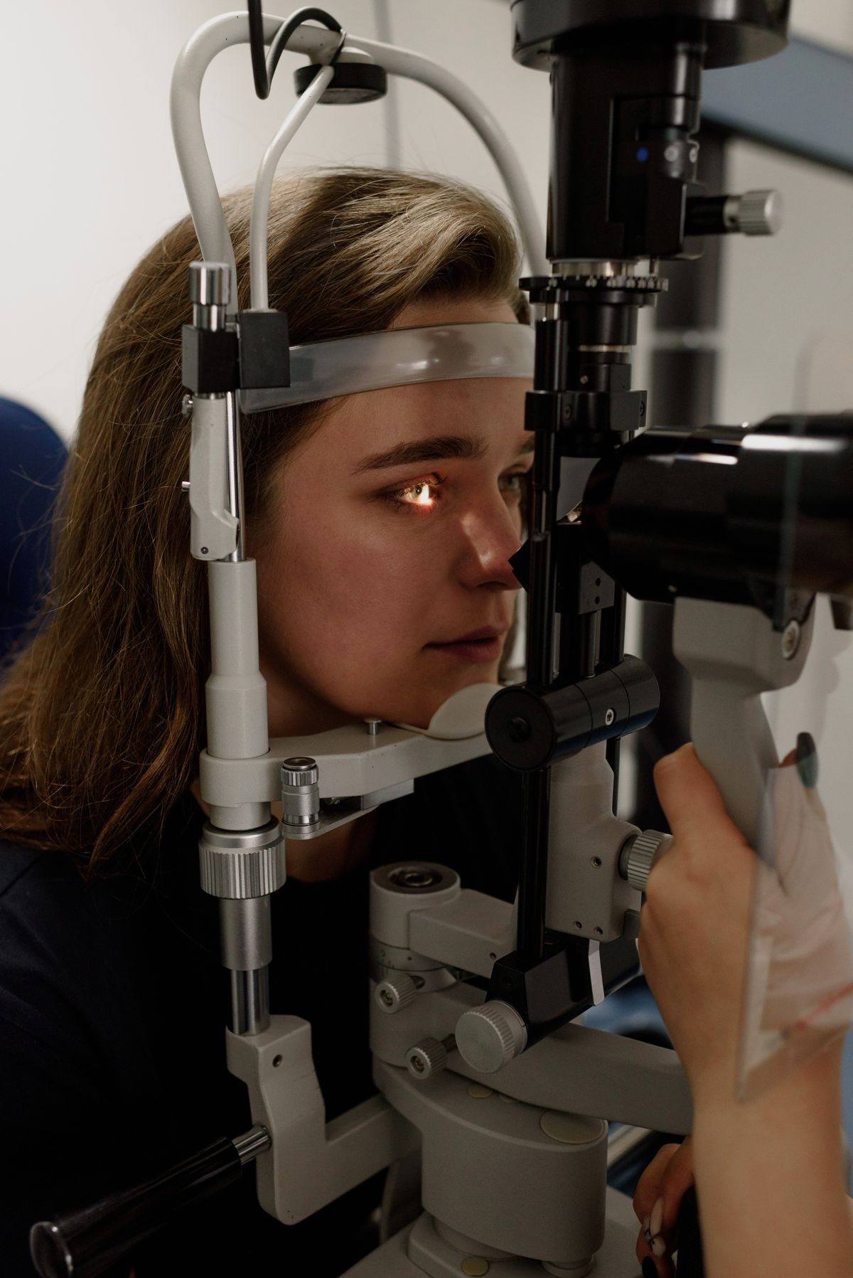
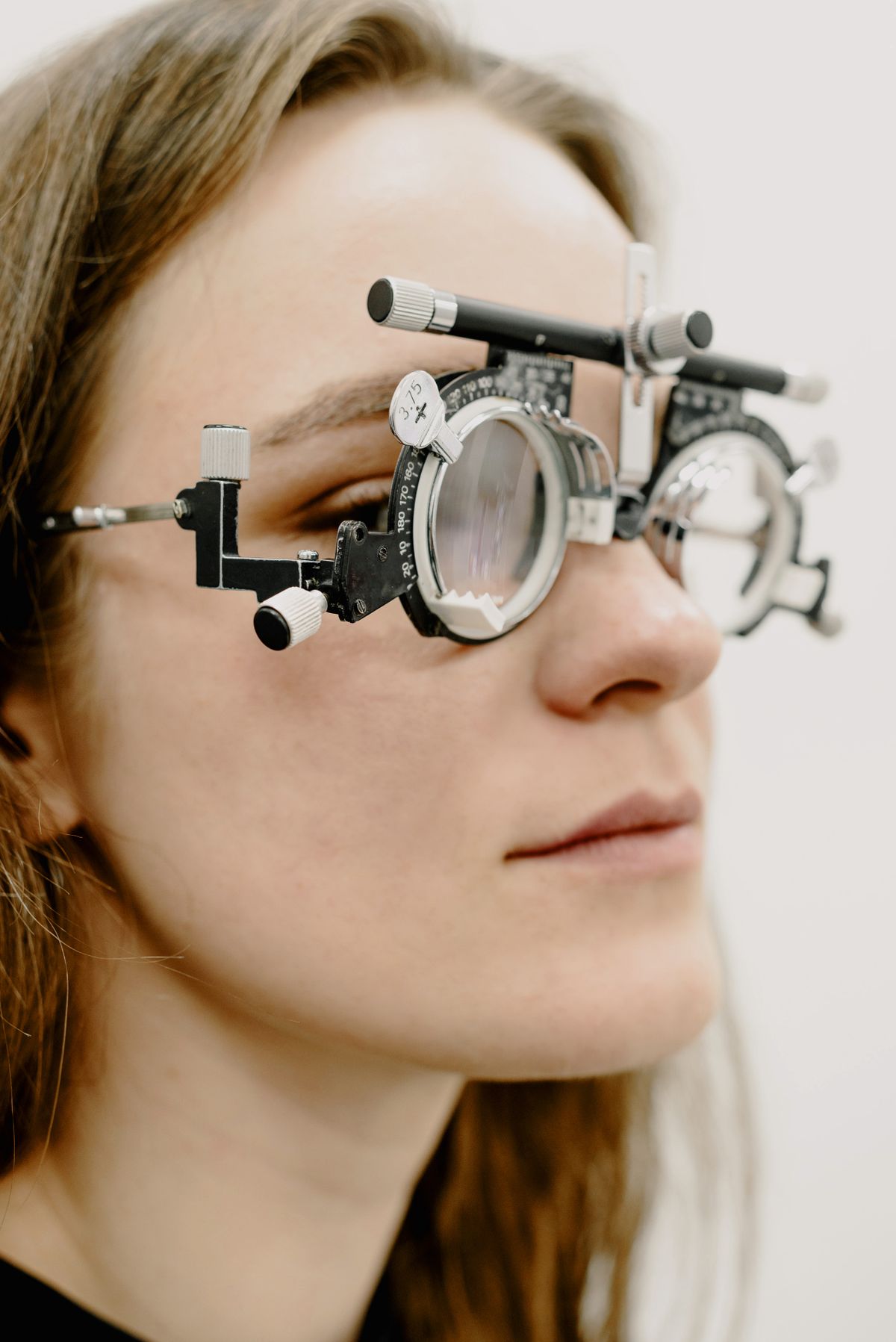
How Birdshot Chorioretinopathy Is Diagnosed
Because birdshot chorioretinopathy is rare, diagnosis is usually made by a uveitis specialist or retina specialist. There is no single “birdshot test,” so doctors combine:
- Detailed history and symptom review
- Comprehensive dilated eye exam to look for the characteristic “birdshot” lesions
- Imaging tests, often including:
- Optical coherence tomography (OCT) – cross‑section pictures of the retina
- Fluorescein angiography – dye test to look at retinal blood flow
- Indocyanine green angiography – highlights choroidal circulation and lesions
- Functional tests such as:
- Visual field testing
- Electroretinography (ERG) to measure how the retina responds to light
- Blood tests to rule out infections or other inflammatory conditions and to check for HLA‑A29
Diagnosis can sometimes take months, especially if early lesions are subtle. Having images and visual fields repeated over time helps the specialist see how things are changing.
“In many patients, the imaging tells the story before the patient fully notices the change. That’s why regular, structured follow‑up is so important in birdshot.”
— Clinical perspective based on current uveitis practice
Current Treatment Options (as of 2026)
The main goal of treatment is to quiet the inflammation and protect vision long‑term, while minimizing medication side effects. Because it is an autoimmune disease, treatment usually focuses on systemic (whole‑body) immune suppression rather than just eye drops.
1. Corticosteroids
Steroids are often used early in treatment or during flares because they work quickly to reduce inflammation.
- Oral steroids (e.g., prednisone) – common at the beginning; doses are tapered as other medications take effect.
- Local injections or implants (around or inside the eye) – for more targeted control, sometimes used if systemic therapy isn’t tolerated.
However, long‑term high‑dose steroid use can cause serious side effects (osteoporosis, weight gain, diabetes, cataracts, glaucoma), so most specialists aim to transition to steroid‑sparing medications.
2. Immunosuppressive “Steroid‑Sparing” Drugs
These medications calm the immune system more broadly and allow steroid doses to be lowered. Common options include:
- Mycophenolate mofetil
- Azathioprine
- Methotrexate
- Cyclosporine (less common now due to side effects but still used in some cases)
These drugs require regular blood tests to monitor liver, kidney function, and blood counts. Many patients stay on them for several years.
3. Biologic Therapies
Over the past decade, biologic drugs have become an important option, especially for difficult‑to‑control birdshot. These are targeted therapies that block specific parts of the immune response.
Biologics used in uveitis (including birdshot) may include:
- TNF‑alpha inhibitors such as adalimumab (FDA‑approved for noninfectious uveitis) and infliximab.
- Other targeted agents in select or research settings, depending on region and evolving evidence.
These drugs are usually given as injections or infusions. They can significantly improve inflammation control for some patients but are expensive and require infection screening and ongoing monitoring.
4. Local Steroid Implants and Injections
In certain situations—such as when systemic drugs aren’t tolerated or when one eye is worse—doctors may recommend:
- Intravitreal steroid injections (temporary effect)
- Long‑acting implants placed inside the eye
These can provide strong local control, but again, they carry risk of cataract and glaucoma. The decision is highly individualized.
Living With Birdshot: Monitoring, Lifestyle, and Daily Life
Managing birdshot chorioretinopathy is a marathon, not a sprint. Even with treatment, most people need years of regular follow‑up.
Ongoing Monitoring
You can expect your specialist to schedule:
- Frequent visits early on (every 1–3 months)
- Ongoing imaging (OCT, angiography) to track inflammation and fluid
- Visual field or ERG testing at intervals
- Bloodwork if you’re on systemic medications
Practical Day‑to‑Day Strategies
While lifestyle changes cannot cure birdshot, they can support overall health and may help you cope better:
- Protect your eyes from glare with good‑quality sunglasses and hats.
- Use high‑contrast, large‑print settings on phones and computers.
- Optimize lighting at home, especially in hallways and stairs, to improve night safety.
- Maintain general health (sleep, balanced diet, physical activity) to better tolerate medications.
- Keep a symptom journal to share patterns and changes with your doctor.

Emotional and Practical Support
It is normal to grieve changes in vision or to feel anxious about the future. Many people find it helpful to:
- Connect with online or local support groups for uveitis or rare eye diseases.
- Speak with a therapist or counselor familiar with chronic illness.
- Inform trusted friends or family so they can support appointments and daily needs.
“One of my birdshot patients said that getting a clear treatment plan and joining a small online community of others with the condition turned everything around for her. She still has to manage flares and medication side effects, but she no longer feels like she’s facing it in the dark.”
— Composite case example based on typical clinical experiences
Prognosis: What Can Patients Expect?
Without treatment, birdshot chorioretinopathy can lead to significant, sometimes severe, visual impairment over time due to damage in the retina and optic nerve. The encouraging news is that earlier and more aggressive treatment has improved outcomes compared with older reports.
Based on recent studies and expert reviews:
- Many patients, especially those diagnosed early and closely followed, can maintain functional vision for years.
- Some may experience permanent blind spots, reduced night vision, or color changes even when central reading vision is preserved.
- Cataracts and glaucoma can develop from both the disease and its treatments, sometimes requiring surgery.

Key Takeaways and Next Steps
Birdshot chorioretinopathy is a rare, serious, but increasingly manageable autoimmune eye disease. While it can’t yet be “cured,” modern treatments give many people the chance to stabilize inflammation and maintain functional vision.
If you or your relative has been diagnosed, you might consider the following steps:
- Confirm care with a uveitis or retina specialist who has experience with birdshot.
- Discuss a long‑term treatment strategy, including steroid‑sparing options and how success will be measured.
- Ask about monitoring plans (OCT, visual fields, bloodwork) and how often you’ll be seen.
- Prepare for lifestyle adjustments to support vision and general health.
- Seek emotional and community support to help navigate the uncertainty and practical challenges.
You don’t have to absorb everything at once. Bring a notebook—or a trusted friend—to appointments, write down your questions, and ask your doctor to explain the plan in plain language. Feeling informed and involved in decision‑making is one of the most powerful tools you have as you move forward with this diagnosis.
Resources and Further Reading
For more detailed, up‑to‑date information, you may find these reputable resources helpful:
- American Academy of Ophthalmology – Eye Health
- U.S. National Institutes of Health – Genetic and Rare Diseases Information Center (GARD)
- Uveitis–related patient education (Massachusetts Eye Research and Surgery Institution)
Because research is ongoing, especially regarding biologic therapies and long‑term outcomes, it’s wise to ask your specialist how the latest evidence applies to your individual situation.
